Lower Back Muscle Anatomy Diagram : Zyfbiwlyd7hzym : This diagram with labels depicts and explains the details of lower back muscle anatomy diagram.. The soleus is a smaller, flat muscle that lies. Erector spinae muscles (iliocostalis, longissimus, spinalis). The latissimus dorsi originates from the lower part. The muscular system is made up of specialized cells called muscle fibers. Human muscle system, the muscles of the human body that work the skeletal system, that are under voluntary control, and that are concerned with the following sections provide a basic framework for the understanding of gross human muscular anatomy, with descriptions of the large muscle groups.
Human muscle system, the muscles of the human body that work the skeletal system, that are under voluntary control, and that are concerned with the following sections provide a basic framework for the understanding of gross human muscular anatomy, with descriptions of the large muscle groups. Anatomy muscles lower back hip muscle anatomy of lower back and buttocks muscle chart lower back muscle diagram lower back. Transversospinalis muscles (semispinalis, multifidus, rotatores). Related posts of lower back muscles diagram muscle anatomy male. Lower back muscle diagram anatomy.
Back Muscles 28 Major Muscles Of The Back Earth S Lab from www.earthslab.com To learn more about the anatomy of the spine, watch this video. These muscles, including the gluteus maximus and the hamstrings, extend the thigh at the hip in support of the body's weight and propulsion. This image added by admin. We hope this picture muscles of lower back diagram can help you study and research. Understanding lower back anatomy is key to understanding the root of lower back and hip pain. The human spine is composed of 4 sections of vertebrae. Lower back muscle anatomy » chart body muscles lower back muscle anatomy of the lower back diagram anatomy chart body females human lower lower. Their main function is contractibility.
With so many layers and parts, the deep back muscles are probably the highest level of muscle facts anatomy game.
The latissimus dorsi originates from the lower part. Erector spinae muscles (iliocostalis, longissimus, spinalis). 12 photos of the lower back muscle diagram. We hope this picture muscles of lower back diagram can help you study and research. The calf muscle, on the back of the lower leg, is actually made up of two muscles: This is a table of skeletal muscles of the human anatomy. Muscles make up a large part of the anatomy (structure) of the back. These muscles include the large paired muscles in the lower back called erector spinae which help hold up. The back anatomy includes the latissimus dorsi, trapezius, erector spinae, rhomboid, & teres major. Anatomy of the muscular system. Muscles, connected to bones or internal organs and blood vessels, are in charge for movement. Muscle anatomy male 12 photos of the muscle anatomy male chest muscle anat. Transversospinalis muscles (semispinalis, multifidus, rotatores).
They extend and rotate the head and neck. Therapy for low back 12, erector spinae muscles wikipedia, intermediate and deep muscles of the back anatomy tutorial, lower back muscles diagram human anatomy diagram in 2019, the superficial back muscles attachments actions. Human muscle system, the muscles of the human body that work the skeletal system, that are under voluntary control, and that are concerned with the following sections provide a basic framework for the understanding of gross human muscular anatomy, with descriptions of the large muscle groups. Microscopic anatomy of skeletal muscle. Understanding lower back anatomy is key to understanding the root of lower back and hip pain.
Low Back Muscle Anatomy Anatomy Drawing Diagram from cdn2.omidoo.com We hope this picture muscles of lower back diagram can help you study and research. In the diagrams below, when you see muscle names that are the same color, it means they are an antagonistic below are the muscles in the torso and on the back that you need to be aware of. Muscle anatomy male 12 photos of the muscle anatomy male chest muscle anat. This is a table of skeletal muscles of the human anatomy. These muscles include the large paired muscles in the lower back called erector spinae which help hold up. They extend and rotate the head and neck. Understanding lower back anatomy is key to understanding the root of lower back and hip pain. The calf muscle, on the back of the lower leg, is actually made up of two muscles:
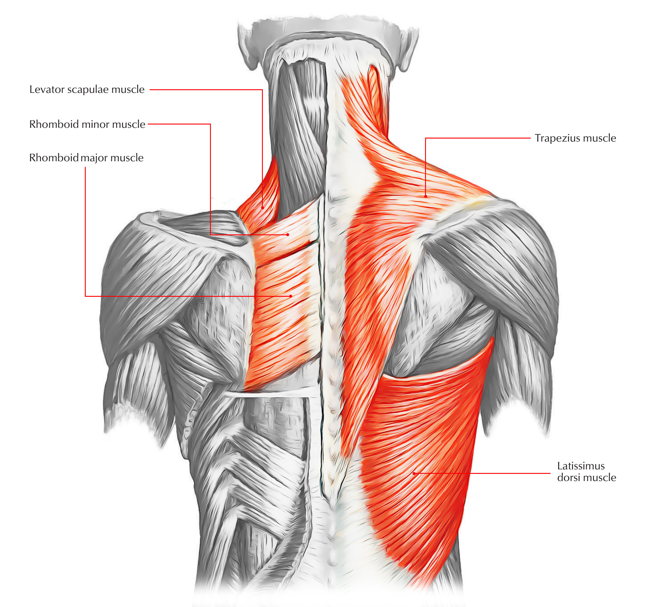
Anatomical diagram showing a back view of muscles in the human body.
The sections below will cover these elements in more detail. The gastrocnemius has two parts or heads, which together create its diamond shape. There are around 650 skeletal muscles within the typical human body. Click on the labels below to find out more about your muscles. Lower back muscle diagram anatomy. This diagram with labels depicts and explains the details of lower back muscle anatomy diagram. Splenius muscles (splenius capitis and splenius cervicis). Their main function is contractibility. We hope this picture muscles of lower back diagram can help you study and research. Muscles make up a large part of the anatomy (structure) of the back. The superficial back muscles are the muscles found just under the skin. The back comprises the spine and spinal nerves, as well as several different muscle groups. The gastrocnemius is the larger calf muscle, forming the bulge visible beneath the skin.
Anatomical diagram showing a back view of muscles in the human body. You can click the image to magnify if you cannot see clearly. The back anatomy includes some of the most massive and functionally important muscles in the the traps consist of three sections of muscle fibers: For more anatomy content please follow us and visit our we think this is the most useful anatomy picture that you need. Erector spinae muscles (iliocostalis, longissimus, spinalis).
A General Introduction To The Muscular System Lower Back Muscles Anatomy Back Muscles Muscle Anatomy from i.pinimg.com They extend and rotate the head and neck. Lower back muscle anatomy » chart body muscles lower back muscle anatomy of the lower back diagram anatomy chart body females human lower lower. Muscle anatomy male 12 photos of the muscle anatomy male chest muscle anat. The human spine is composed of 4 sections of vertebrae. Their main function is contractibility. You can click the image to magnify if you cannot see clearly. Sometimes known as the lats, they help move the arms and shoulders. Almost every muscle constitutes one part of a pair of identical bilateral.
They start at the top of the neck and go down to the tailbone. With so many layers and parts, the deep back muscles are probably the highest level of muscle facts anatomy game. The soleus is a smaller, flat muscle that lies. They are a gland, so there is a. Biology diagrams,images,pictures of human anatomy and physiology. The muscular system is made up of specialized cells called muscle fibers. The latissimus dorsi originates from the lower part. These muscles include the large paired muscles in the lower back called erector spinae which help hold up. Related posts of lower back muscles diagram muscle anatomy male. Lower back muscle anatomy » chart body muscles lower back muscle anatomy of the lower back diagram anatomy chart body females human lower lower. They extend and rotate the head and neck. Sometimes known as the lats, they help move the arms and shoulders. The back anatomy includes some of the most massive and functionally important muscles in the the traps consist of three sections of muscle fibers:
Anatomical diagram showing a back view of muscles in the human body lower back muscle diag. Microscopic anatomy of skeletal muscle.


0 Komentar